![]()

There are many uses in the world of dogs and cats for Ultrasound equipment, I specialise in two areas
-
Pregnancy Detection
-
Post whelping to ensure all whelps or kits are delivered
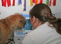
An ultrasound machine emits ultrasound waves. They are reflected back into the hand-held probe that is placed on the skin. The pattern of the reflected sound waves creates an image that is viewed on a screen. It is non invasive and safe , there is no need to anaethetise the bitch or queen and because the owner can stay close she can be reassured and held normally. To assist in better picture the use of a conductive gel is sometimes used and a small amount of hair in heavily coated breeds may be clipped away.
Pregnancy detection can be carried out from 25 days, I have found that 28 days gives a clearer and more accurate reading. As the pregnancy progresses it becomes more difficult to assess numbers.
During the scan I can capture and print a photograph of the puppies or kittens safely nestled in the bitches womb, a lasting momento of a great moment
Cost: £25.00 (photograph included)
Travel and journey time costs will be calculated on .60p per mile, single journey basis.
Equipment is mobile and can be used within your own home thus causing less distress to the bitch or queen and viable babies
Diagnostic Ultrasound in Canine Reproduction
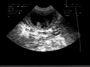
The human ear can detect sound waves of certain frequencies; higher frequencies are outside this range. These higher frequency sound waves may be directed into a small beam of sound; the principle of diagnostic ultrasound. The ultrasound beam is produced by small crystals which are housed within a transducer or probe. These crystals both produce the sound (by expanding or contracting when an electric current is applied to them) and receive the returning sound beam and convert it back into an electrical signal. The electrical impulses produced by the returning sound are then converted into a picture on a small screen. The picture produced on the screen is referred to as an image7. The ultrasound crystals produce many rapid pulses of ultrasound which allow images to be continually updated on the screen so that movement of organs can be seen. This is called real-time ultrasonography and is now the most commonly used method.
When a beam of ultrasound is directed into the body from an ultrasound machine the sound travels through tissue until it meets an obstruction. At this point some sound continues on its original path, whilst some is reflected back to the ultrasound machine. The speed of sound within the body is almost constant; therefore by measuring the time taken for the echo to return to the machine, the distance between the ultrasound machine and the tissue obstruction can be calculated. This calculation is done by complicated electronics within the machine and the information is displayed on the ultrasound screen. Ultrasound pictures are black and white with varying shades of grey. The brightness of the image is related to the degree of sound reflection. This type of machinery is called brightness (or B-mode) ultrasound. If most of the ultrasound beam is reflected back to the transducer the images appear white on the screen. When little of the sound is reflected the images appear darker shades of grey or black. Very dense structures such as bone reflect nearly all of the sound beam and produce bright white images. Gases such as air (found in the lungs and in some areas of the gastrointestinal tract) do not allow the transmission of ultrasound and also appear white on the ultrasound screen. Conversely most fluids allow sound to be easily transmitted; they appear black on the ultrasound screen. The amount of fluid within an organ therefore affects its appearance when it is examined with ultrasound. The pictures produced by the ultrasound machine may be observed during the examination. However, it is often best to save images on a video recorder so that they can be reviewed later, or to take copies of images using a polaroid camera or video printer.
Ultrasound may be used to image the uterus of most bitches. This is most useful for the diagnosis of pregnancy, the assessment of foetal numbers and the assessment of foetal viability
There are many reasons why bitches may be examined by ultrasound to confirm pregnancy. It may be important to diagnose pregnancy relatively early to allow adjustments to be made in feeding and routine medication regimes and is an important consideration if therapeutic drugs are to be used. Ultrasound examination may be undertaken in mid-pregnancy in bitches that were not examined earlier or where there is concern over foetal resorption. Ultrasound may be used when it is necessary to differentiate between pregnancy and pyometra since in mid-pregnancy bitches frequently have a vaginal discharge and refuse their food.
In late pregnancy bitches may be examined if they are thought to be 'overdue' or if there is concern over foetal abnormalities. Bitches which have problems during whelping may be examined to confirm whether the pups are still alive, and bitches may be examined after whelping to ensure that no pups have been retained. There is no evidence to suggest that ultrasound examination is harmful to the developing pups.
There is some confusion about when ultrasound can be used to diagnose pregnancy in the hitch. This is not surprising considering the complicated events which occur around the time of ovulation. The majority of bitches whelp 63 days after ovulation. Some bitches may stand and be mated several days before ovulation. If sperm from an 'early mating' live long enough to fertilise eggs, the time from mating to whelping will be longer than 63 days, and can be up to 72 days.
Some bitches, however, will stand and be mated several days after ovulation. If eggs are still present during a 'late mating' these may be fertilised. In this case the time from mating to whelping will be shorter than 63 days, and can be as little as 58 days. Therefore, although the time from ovulation to whelping is nearly the same in all hitches, the time from mating to whelping (the apparent pregnancy length) can vary between 58 and 72 days.
This has important implications for the early diagnosis of pregnancy in the bitch. The size of the foetus is related to the timing of ovulation and not the timing of mating. Bitches which have been mated 'early' may be presented for ultrasound examination too soon, before foetal fluids have accumulated. These bitches may therefore be thought to be non-pregnant. Pregnancy can be detected using ultrasound as early as 19 to 22 days after ovulation. Ovulation time in most bitches is not known so that 28 days after the last mating is a suitable time to conduct an ultrasound examination.
In early pregnancy the embryo is present within a small amount of fluid. The embryo, which is only 1-2 mm in diameter, appears grey on the ultrasound screen and is suspended within a fluid filled swelling (black cavity) of the uterus. A small flickering of the embryonic mass (representing the beating of the heart) can often be seen. As the embryo grows it becomes possible to recognise distinct head and body regions, and the foetal membranes become obvious. The foetal sac enlarges and the embryonic tissues become more prominent and the heart is more easily seen.
At an early stage of pregnancy (28 - 35 days) it is possible to estimate the number of pups. This requires careful examination, and can be difficult even for experienced operators. It is often easiest when there are only one or two pups and is more difficult in later pregnancy when only sections of each pup are seen. The accuracy in determining the actual number decreases with larger litters; the overall accuracy is low (about 30%). Most frequently the number of pups is underestimated.
In mid-pregnancy (35 - 55 days) the foetal skeleton develops and can be seen. This appears white on the ultrasound screen. The head appears circular early on but later the jaws and nasal bones can be identified. The spinal column, pelvis and the ribs can also be seen. The internal organs become more prominent in their appearance. The liver is in the front of the abdomen, being dark grey in appearance, and the fluid filled stomach is often noted just behind this. The heart and some of the great arteries and veins can also be identified. Movements of the pups are often noted and individual limbs can often be seen.
In late pregnancy the bladder, which is fluid filled, may be seen. The other abdominal organs become more prominent and look more like those of a mature animal. The chambers of the heart can easily be identified and organs like the kidneys are more easily seen. Counting the number of pups at this time is less accurate than earlier. Pups which are dead can he identified by an absence of heart beats and a lack of movement and foetal fluid.
Most ultrasound machines possess electronic callipers which can measure the structures being examined. A variety of measurements can be made on individual pups. Measurements of the head or thorax size for example can be compared to normal tables, thus allowing an estimation of the age of the pups to be made. This allows the time of whelping to be predicted. This procedure is commonly applied to pregnant women but to date has only been done in one breed of dog (the labrador retriever). Since there is such a large size difference between breeds normal values for one breed are of little use to others.
Abnormalities of pregnancy
Regular ultrasound monitoring of bitches has demonstrated that there is probably a higher incidence of resorption than is generally appreciated. This information is interesting but has limited value since in most cases little can be done to prevent resorption once it has started. Indeed it may be dangerous to attempt this since in many cases there is a good reason for the resorption. Foetal distress and death may be noted with ultrasound. This may help to decide whether to perform a caesarean operation when assessing a bitch with a prolonged whelping. If dead pups are present, gas may be detected within the uterus. In man, many foetal abnormalities can be detected using ultrasound. This is helped by the large size of the foetus and the fact that single foetuses are most common. In the dog it is difficult to examine all foetuses in mid and late pregnancy.